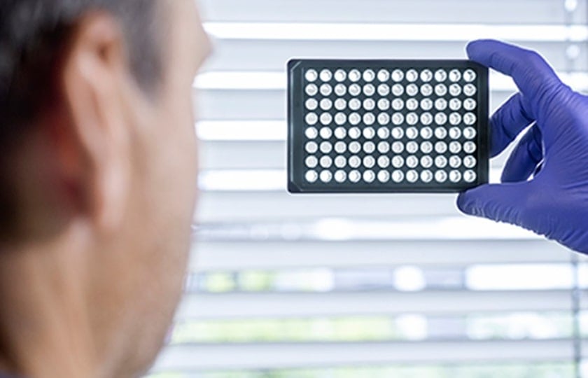
With low autofluorescence, high optical properties and high transmission in the low UV range, SCREENSTAR is a highly suitable microtiter plate for classical high throughput and high-content screening (HCS) applications, or whenever high-quality imaging is critical to results. [1,2]
Why SCREENSTAR microtiter plates provide exceptional imaging properties for HCS
Plate material
SCREENSTAR microtiter plates are made entirely of cycloolefin. They have a black pigmented frame with a 190 µm thick ultra-clear film bottom, showing both excellent optical properties comparable to those of glass and surface modifications optimized for cell culture.

Why cycloolefins are so versatile
Cycloolefins are an excellent base material for microtiter plates because they combine many useful properties, including high glass transition temperature, optical clarity, a low autofluorescence background in the lower UV range, low shrinkage, low moisture absorption and low birefringence.[1] The material is also ideal for storage of substances due to its low water absorption, high vapor barrier, low levels of leachables and resistance to DMSO, which is the most commonly used solvent in high-throughput screening. Cycloolefins have excellent optical properties with a refractive index and focal background comparable to glass.
Optics and image quality
With highest optical transparency, low autofluorescence and absorption in the lower UV range, low birefringence and a refractive index of 1.53 SCREENSTAR microtiter plates offer superior data quality in biochemical assays. With a bottom thickness of 190 µm, the plates are designed to meet the optical requirements of most microscopic systems for excellent image quality with high resolution – in contrast to other products, whose thicker bottoms are outside the tolerance ranges and thus compromise optical resolution – an all-important factor in, for example, signal counting-based assays.

Geometry
Geometry and material properties make SCREENSTAR microtiter plates ideally suited for fully automated screening systems. Due to the recessed bottom of the wells, it is possible to use oil- or water-immersion objectives with high magnification to gain access to all wells of the microtiter plate without limitations in edge and corner positions. SCREENSTAR microtiter plates are available in 1536 well, 384 well and 96 well formats. Dimensions and tolerances of the plates comply with the respective ANSI standards. To improve cell culture conditions and reduce evaporation, the 96 well SCREENSTAR microtiter plates have a ditch around the perimeter which can be filled with sterile media or water to help create a saturated water vapor barrier.
Surface chemistry
Homogeneous cell attachment and growth is critical for reliable results in cell based high-content or high-throughput screening. SCREENSTAR microplates are available both sterile with a hydrophilic, cell culture-treated surface for cell-based assays and untreated with a hydrophobic surface for biochemical assays. In addition, they can also be provided with biological coatings, Advanced tissue culture (TC) modification or high-binding treatment. Please feel free to contact us directly if you need more information or application support.
Do you want to learn more about microtiter plates for advanced microscopy in high-content and high-throughput screening? Then have a look at our website.
References
[1] SCREENSTAR: A 1536 Well Microplate for High Content and High-Throughput Screening. Greiner Bio-One forum – Technical Notes and Applications for Laboratory Work No. 15 https://www.gbo.com/fileadmin/media/GBO-International/01_Downloads_BioScience/SALES_Scientific_Publications/F073120_Forum_No._15_SCREENSTAR.pdf
[2] SCREENSTAR and CELLview: Microplates for Advanced Microscopy. Greiner Bio-One forum – Technical Notes and Applications for Laboratory Work No. 18 https://www.gbo.com/fileadmin/media/GBO-International/01_Downloads_BioScience/SALES_Scientific_Publications/F073787_Forum_No._18_SCREENSTAR_CELLview.pdf