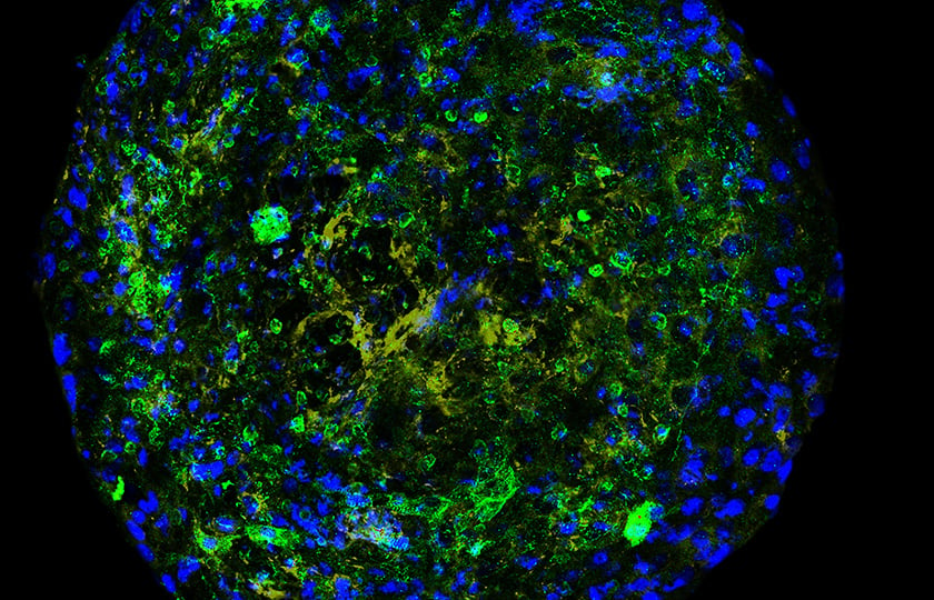
Organoids and Spheroids in 3D Cell Culture
Spheroids and organoids – do you know the difference?
Organoids and spheroids are often used as interchangeable terms, especially by people new to the field of three-dimensional (3D) tissue modeling. One might get the impression that both terms mean the same thing, and you could be forgiven for thinking so. However, this is not always accurate. Understanding the difference is not that complicated, because the meaning of organoids and spheroids is quite intuitive.
Spheroids are spherical cellular entities that are generally grown as free-floating or suspended aggregates. They are usually grown from immortalized cell lines or fragments of human tissue. The cells are grown to adhere to each other rather than to a growth medium or the solid walls of a container.
Organoids, on the other hand, can be described as 3D structures that are grown primarily from single stem cells, often clonogenic, that differentiate into structural units with the tissue organization of an organ. In contrast, spheroids are formed by aggregating cells of different or the same type to achieve one or many organotypic phenotypes. Organoids been grown from many tissues such as intestine, retina, brain, liver, lung, and kidney.
In summary, organoids and spheroids are generated to better mimic the morphology and physiology of in vivo tissue as an in vitro model. The best choice of 3D model will always depend on the hypothesis interrogated and the level of simplicity or complexity of the 3D model required to execute a project.
Is there a key difference between organoids and spheroids?
The crucial difference between organoids and spheroids is the driving force behind aggregation. In spheroids, cell-cell adhesion mediated by proteins is responsible for their stability. On the other hand, the cells in organoids are connected during the co-developmental processes, when they are generated through stem cell differentiation.
What are the individual advantages and disadvantages?
Both organoids and spheroids are popular tools for studying the response of tissues to external factors such as drugs, physical stimuli, or pathogens. For example, a recent study using spheroids derived from bronchial epithelial cells examined how environmental parameters such as air pollution affect our airways [1]. Organoids, depending on the type of tissue, can more closely mimic the cellular environment in vivo tissue. However, it can be difficult to obtain organoids because their phenotype and viability depend highly on the media and matrix that support their development.
Both spheroids and organoids are also commonly used in the study of tumors, especially in personalized medicine. Cells can be taken from a cancer patient, which can then be grown into a 3D cell model of that specific cancer. Because a cancer is a genetically unique entity, it responds individually to specific drugs and treatments. 3D cell models can be used to test the sensitivity of a cancer to help guide patient treatment. Both spheroids and organoids can be used to study the microenvironment and model the macroscopic features of a tumor.
When should you use organoids, when spheroids?
Which model to choose for a particular study must be carefully selected depending on the cell type, the planned experiments, time, and budget constraints. Spheroids, for example, are generally not only faster to form but also easier to maintain. Organoids often require specific cofactors to induce differentiation and specific media conditions for culture viability; therefore, they can be impractical and expensive. Organoid phenotype highly depends on the media or matrix used to culture them. The matrices used to grow organoids are often derived from animal tissue. Therefore, there is significant batch-to-batch variability in the production of these reagents that are known to impact the reproducibility accuracy of these models significantly.
How do you produce organoids and spheroids?
When attempting to create custom 3D cell models, there are a variety of different methods. The most important distinction is whether a method is scaffold-based or scaffold-free. Spheroids can be grown using either method. Scaffold-free methods are generally simple and fast. For example, spheroids can be generated by simply centrifuging at the correct speed [2]. Another method for generating spheroids is the hanging drop method. At the bottom of the droplet, cells begin to adhere to each other due to surface tension and gravity, forming a spheroid.
Interestingly, it is also possible to form spheroids using magnetic forces. To do this, the cells need to be magnetized, e.g., using the Greiner Bio-One Nanoshuttle-PL in overnight culture [2]. The cells treated in this way can be seeded onto a plate with magnets, leading to their directed assembly above the magnet and promoting fast cell-cell interaction leading to expedited spheroid formation.
Scaffold-based approaches attempt to mimic the natural environment and shape of cells by providing a matrix in which cells can grow. For this reason, scaffold-based approaches are mostly used to grow organoids. Some commercially available substances used for this purpose are derived from animal basement membranes and contain a variety of proteins important for signal transduction from the extracellular matrix to cells and can provide important stimuli, especially for epithelial cells that normally rest on basement membranes in vivo. However, matrices that are produced directly from animals require significant amounts of tissue for mass production, which is ethically questionable and a source of batch-to-batch variability, known to hamper the viability and reproducibility of these models. 3D cultures using magnetic 3D (M3D) cell culture have shown to produce more complex and in vivo sphere-like extracellular matrices than other commercially available products [3].
If you are interested in research with organoids and spheroids, you can learn how scientists can use 3D models to facilitate drug discovery and fight disease on 3D Made Easy, our site for all things in 3D cell culture technology.
Ready to enter the next level?
Please fill out this form and contact our experts today to find the perfect solution for you!
Don't miss our regular updates on scientific topics around 3D Cell Culture
References
[1] Baarsma HA, Van der Veen CHTJ, Lobee D, Mones N, Oosterhout E, Cattani-Cavalieri I, Schmidt M. Epithelial 3D-spheroids as a tool to study air pollutant-induced lung pathology. SLAS Discov. 2022 Apr;27(3):185-190. doi: 10.1016/j.slasd.2022.02.001. Epub 2022 Feb 25. PMID: 35227934.
[2] Bosnakovski D, Mizuno M, Kim G, Ishiguro T, Okumura M, Iwanaga T, Kadosawa T, Fujinaga T. Chondrogenic differentiation of bovine bone marrow mesenchymal stem cells in pellet cultural system. Exp Hematol. 2004 May;32(5):502-9. doi: 10.1016/j.exphem.2004.02.009. PMID: 15145219.
[3] Vu B, Souza GR, Dengjel J. Scaffold-free 3D cell culture of primary skin fibroblasts induces profound changes of the matrisome. Matrix Biol Plus. 2021 May 12;11:100066. doi: 10.1016/j.mbplus.2021.100066. PMID: 34435183; PMCID: PMC8377039.
