
In cell-based screening, the tissue culture surface plays a decisive role in the cell’s behavior and hence the reliability of the screening results. The right protein-coated surface can enhance cell adhesion and provide the environmental cues essential for maintaining disease-relevance of primary cells and other challenging model systems. Selecting the ideal protein coating is not always straightforward, but fortunately help is at hand.
Mimicking the extracellular matrix in vitro
In their natural environment, cells surround themselves with an extracellular matrix (ECM) that provides structural support and mediates cell signaling pathways important for maintaining their phenotypic and functional characteristics. The composition of the ECM differs from one tissue to another, and subtle differences can profoundly influence cell adhesion, morphology, gene expression and many other characteristic behaviors.
Each tissue-specific ECM comprises a characteristic assortment of structural and adhesive proteins, including various isoforms of collagen, laminin, and fibronectin. With a vast selection of ECM components and extracts available commercially, assay developers have the power to tailor the culture environment to mimic a particular ECM and optimize assay performance.
However, sourcing and testing the effect of different coatings and protein combinations can be a laborious, time-consuming and cost-intensive exercise.
Overview of peptide & protein coatings
- Poly-D-Lysine (PDL) & Poly-L-Lysine (PLL) are synthetic poly-peptides commonly used for cell culture coatings, providing a highly positive charged surface that enhances cell adhesion. PDL coatings are more durable based on their resistance against proteases released by certain cell types.
- Collagen Type I (Col. Type I) is a major component of the extracellular matrix (ECM) of numerous cell types. Collagen coatings help mimic the in-vivo microenvironment and promote cell adhesion, proliferation and differentiation of mesenchymal and connective tissue-derived cells.
- Fibronectin is a glycoprotein that connects membrane-spanning receptors (“integrins”) to extracellular matrix proteins like collagens and heparan sulfate proteoglycans. Fibronectin-binding integrin receptors are present in cell membranes of numerous cell types making fibronectin coatings a preferred choice to facilitate fast, strong attachment, spreading and growth of many cell types.
- Laminin is a glycoprotein and major component of the basement membrane. It binds to integrin cell surface receptors in the cell membrane and to type IV collagen, fibronectin and proteoglycans in the extracellular matrix. As a vital support for scaffolding the basal lamina, laminin coatings promote structural integrity, cell adhesion, migration and tissue organisation, and are commonly used for culturing neurons, muscle cells, epithelial and endothelial cells.
- Basement membrane extract (BME) is derived from Engelbreth-Holm-Swarm mouse sarcoma, composed predominantly of laminin, type IV collagen and heparan sulfate proteoglycan. It mimics the basement membrane extracellular environment in many tissues and is used as a “thin coating” for cell adhesion and support of cell-specific phenotype in complex 2D-cell models.
A fast route to success
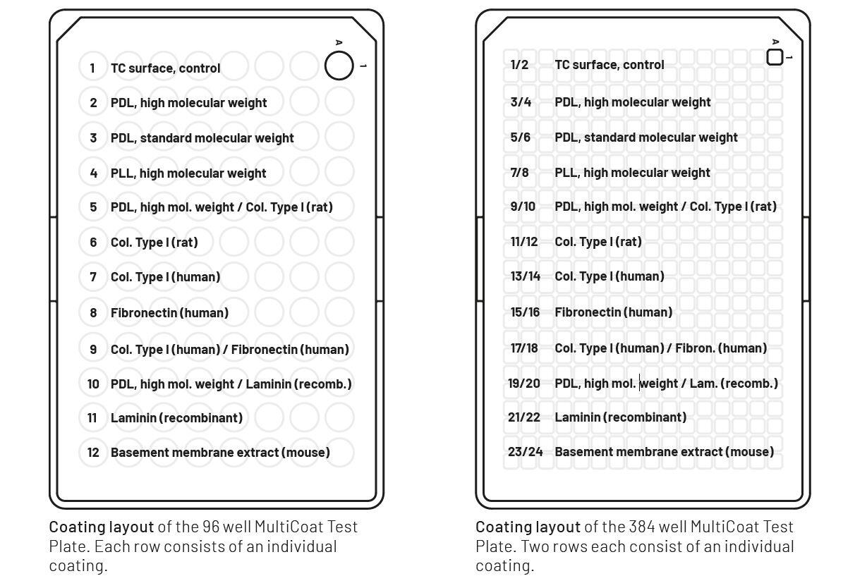
To make testing easier and more cost-effective, Greiner Bio-One has introduced a new MultiCoat test plate, which offers a choice of 11 different single and double coatings on a premium imaging plate in 96-well or 384-well format.
The multicoat test plate can be used to rapidly screen a variety of coatings at once to find the appropriate biological environment for the cells. It’s an optimal tool to find the adequate cell culture conditions in the early phase of the development of cell-based assays, or for further development of existing assays to optimize critical factors such as cell adhesion, distribution, morphology and differentiation.
After pre-screening different coatings with the multicoat test plate, you can confidently transition to high throughput or high-content screening (HTS/HCS) using SCREENSTAR CELLCOAT® 96, 384 and 1536 well microplates pre-treated with the optimal coating of your choice.
Finding the right protein-coated surface just got easier
The new MultiCoat test plate makes finding the right coating for your cell-based assay quicker, easier, and more cost-efficient than ever before. If needed, our experts are on hand to offer additional support in choosing the ideal coating for your specific application.