Selecting an optimal microtiter plate for an assay can be difficult because there are multiple options and competing tradeoffs in microtiter plate selection at each step of the assay development process. There are both application-specific aspects of choosing the right microtiter plate and technique-specific requirements. In this article, we will look at important considerations for various high-throughput screening assays and compound management.
Microplate Selection Guide

Not all microtiter plates are created equal: technical considerations
Surface properties of microtiter plates play an important role for the functionality and can be modified physically, chemically or with coating methods to fulfill various demands.
Cell-free or cell-based assays?
Microtiter plates for cell-based assays are usually treated and sterilized for tissue culture and may have clear bottom wells for visualization and special coatings to enhance or prevent cell attachment. For cell-based assays, you need to ensure that your cells grow in a way that most closely resembles the in vivo state. Microtiter plates for cell-free assays may have a range of surface conditions, from non-binding surfaces to minimize protein adsorption (e.g., enzymatic homogeneous assays), to strongly binding surfaces that enhance adsorption (e.g., ELISA). [1]
Homogenous or heterogenous assays?
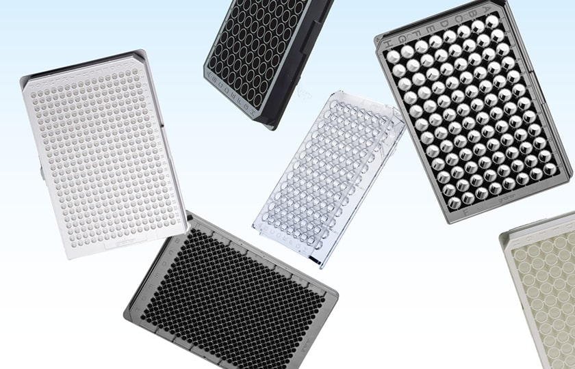
For homogeneous assays, minimal binding to the surface is desired, while heterogeneous assays such as ELISA [2,3] require adsorption of molecules or cells to the bottom of the microtiter plate. Depending on the type of ELISA you want to perform, whether you are analyzing whole cells, and depending on the method you choose for quantification, e.g., colorimetric or by luminescence/fluorescence, you will use different microtiter plates with appropriate surface properties (e.g., binding/non-binding, transparent/nontransparent). Black pigmented microtiter plates are often used for fluorescence applications, while white pigmented microtiter plates typically support (chemi)luminescence measurements and are sometimes used to amplify fluorescence signal intensity.
Microscopy, imaging, and high-content screening
Applications in high-content screening (HCS), as well as high resolution and confocal microscopy, require microtiter plates with pigmented walls and a clear bottom. They are a prerequisite for luminescence and fluorescence applications where bottom reading or microscopy are involved. HCS applications generally require high-quality images and involve an analysis of whole cells or cell components with simultaneous readout of multiple parameters. Therefore, HCS requires a sophisticated readout process; microscopy often in conjunction with image recognition and AI-based algorithms. Such imaged-based assays place high optical requirements on the microtiter plates used to yield the best possible result.
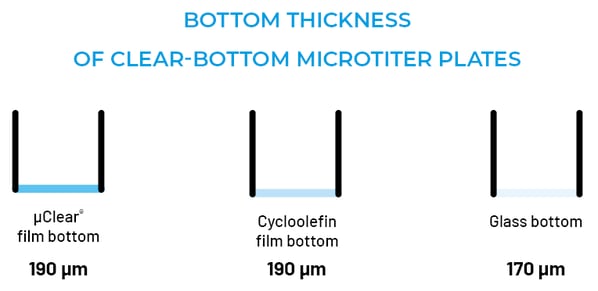
The ideal microtiter plate bottom surface should be uniform with a bottom thickness of approximately 170 (± 20) µm to match microscope objective lenses with high numerical apertures. Greiner Bio-One´s glass-bottom microtiter plates have a bottom with a thickness of 170 µm and show excellent uniformity for both inter- and intra-well flatness over the entire size of the plate, which makes them the perfect choice for demanding applications requiring the highest optical quality. Glass-bottom microtiter plates may be more expensive than microtiter plates made of other materials due to the complex manufacturing process, but if you need the best imaging quality for HCS, it can be worth it. [4]
Microtiter plates with cycloolefin film bottoms are optimized for the specialized requirements of high-content screening and high-resolution microscopy. The cycloolefin film bottom guarantees maximum resolution, even at high microscopic magnification, and the physical surface treatment assures a proven performance for consistent cell attachment.
Permeable membrane inserts for functional polarized cells
To perform whole cell polarization, migration and differentiation experiments, permeable membrane supports are an excellent method. You can read in detail about the advantages and applications of permeable supports in one of our next blog series.
Assay miniaturization
Miniaturization of assays plays a major role especially in high-throughput screening. Usually this is done by changing from a 96 well plate to a 384 well plate or even higher format. However, miniaturization often comes with crucial changes in handling of the plates and adaptation of the laboratory instrumentation. 384 well plates are considerably more expensive than 96 well plates but reducing the sample volume can be an important aspect to save expensive reagents and cells and increase throughput. An alternative to the use of high-format microtiter plates are Half Area microtiter plates, which allow a reduction of the sample volume by up to 50%. Small Volume HiBase microtiter plates are another option for assay miniaturization. These plates allow top reading even at low working volumes and reagent savings similar to 1536 well microtiter plates. They are suitable for transmission, fluorescence and luminescence applications and offer excellent optical properties. [5] For more info on assay miniaturization, see our corresponding blog series here.
Well characteristics – density, shape, well volume
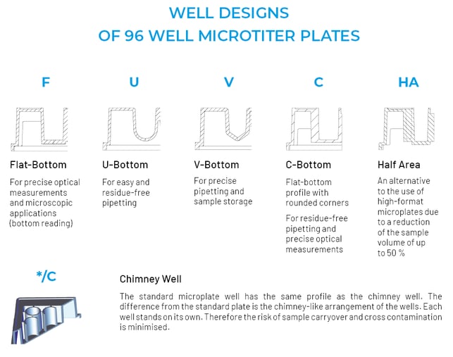
Microtiter plate well geometries typically are round, square, or rounded-square and follow the choice of assay. The individual geometries have specific advantages. Round wells have a smaller total area compared to square wells, minimizing reaction volume. Square wells provide maximum area for light transmission. Flat bottoms are ideal for bottom-reading applications, and flat bottom chimney wells are used to reduce cross contamination. Conical bottoms (V-bottoms) allow maximum removal of small sample volumes. Rounded/spherical bottoms (U-bottoms) facilitate mixing, washing, coating, and removal of solutions from wells. [1]
Long-term storage, sample, and compound management
Storage plates are biologically inert, resistant to temperature changes and solvents such as DMSO. Raised well walls facilitate sealing. Suitable covers are available for the long-term storage of microtiter plates. A sterile and tight seal prevents contamination and sample evaporation. Greiner Bio-One also offers special storage plates available from 96 to 1536 well plate formats and with U- and V-bottom designs.
Would you like further assistance in selecting the optimal microtiter plate for your high-throughput screening application?
Microplate Selection Guide
References
[1] Auld DS Ph.D., Coassin PA B.S., Coussens NP Ph.D., et al. Microplate Selection and Recommended Practices in High-throughput Screening and Quantitative Biology. In: Markossian S, Grossman A, Brimacombe K, et al., eds. Assay Guidance Manual. Bethesda (MD): Eli Lilly & Company and the National Center for Advancing Translational Sciences; June 1, 2020. https://pubmed.ncbi.nlm.nih.gov/32520474/
[2] Microplates for Enzyme Linked Immunosorbent Assays (ELISA). Greiner Bio-One forum – Technical Notes and Applications for Laboratory Work https://www.gbo.com/fileadmin/media/USA/01_Downloads_BioScience/SALES_Scientific_Publications/F073004_Forum_No._09_ELISA.pdf
[3] Insulin ELISA on high binding MICROLON ® 600 and CELLSTAR ® Microplates. Greiner Bio One Application Note https://www.gbo.com/fileadmin/media/USA/01_Downloads_BioScience/SALES_Scientific_Publications/F073106_AN_InsulinELISA.pdf
[4] Plastic Labware for Optimal Results in Modern Life Science Microscopy Using ZEISS Axio Observer and ZEISS Celldiscoverer 7. Technology Note. Philipp Wachter (Greiner Bio-One GmbH, Frickenhausen, Germany), Martin Gleisner, Volker Doering (Carl Zeiss Microscopy GmbH, Jena, Germany). February 2022 https://asset-downloads.zeiss.com/catalogs/download/mic/98a1fce0-9d82-4d0b-b95c-dc3ffcbe1376/EN_wp_GBO_microscopy_grade_labware_on_ZEISS_microscopes_.pdf
[5] Greiner Bio-One Microplate Selection Guide https://www.gbo.com/fileadmin/media/GBO-International/01_Downloads_BioScience/SALES_Brochures/English/F073048_microtiter plate _Selection_Guide_EN.pdf