High-Throughput Screening and Beyond with Magnetic 3D Bioprinting
Magnetic 3D bioprinting (M3D) combined with Cell-Repellent surfaces
shows promising results with HTS for evaluating new chemotherapeutics to fight pancreatic cancer.
Content of this article:
- Faster, cheaper, and better high-throughput screening (HTS)
- How to choose your HTS assay
- Why Magnetic 3D Bioprinting with HTS?
- Expert-Talk with Dr. Timothy Spicer about his research using HTS combined with m3D cell culture technology for drug discovery lead identification against ex vivo pancreatic tumor models
Faster, cheaper, and better high-throughput screening (HTS)
Cell-based HTS aims to select leads or hits from compound library screenings that elicit an intended cell response. This is done with automation/robotics, plate readers, high-content imagers, and dedicated instrumentation control and data processing software, such as artificial intelligence (AI). The time and cost associated with each step can vary with the targeted biomarker, readout, and data analysis approach. HTS can take from one week (1,000 to 10,000 compounds) to months (10,000 to millions of compounds). For improving predictiveness and reducing false positives of HTS compound screening[1], M3D has shown to be an effective and superior tool for achieving the desired tissue phenotypes[2][3] [4][5] needed to power faster, cheaper, and better results:
| Faster | Cheaper | Better |
|
|
|
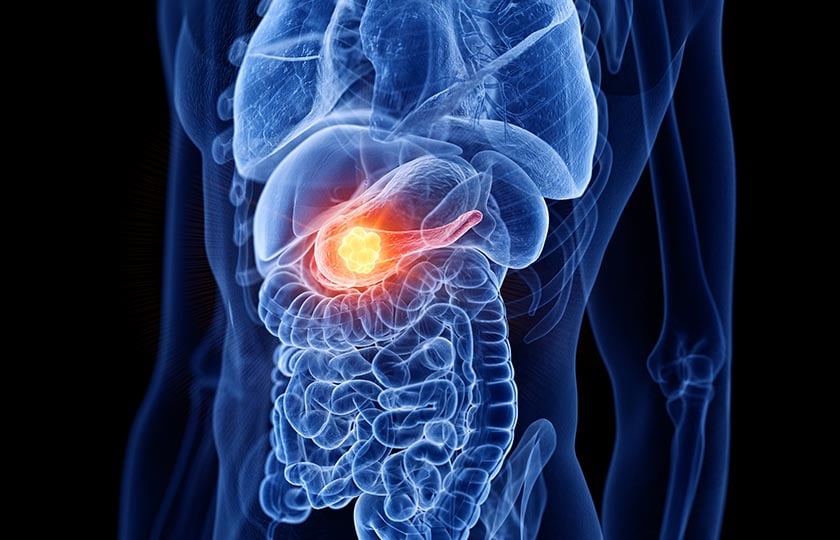
Three dimension cell culture models have become essential for evaluating new chemotherapeutics to fight pancreatic cancer.
Three dimension cell culture models have become essential for evaluating new chemotherapeutics to fight pancreatic cancer. The research by Fernandez-Vega et al.[2] screened 150K small molecule library using M3D and cell-repellent microplates (384 and 1536 well) against primary pancreatic organoids; this was the first report of an HTS of this scale using clinically relevant pancreatic tumor model generated from patient biopsies.
Magnetic 3D Bioprinting (M3D) enables the uniform generation and ease manipulation of spheroids/organoids for HTS by directing magnetized cells to assemble in a controllable size within a shorter culture time. Thus, this technology surpasses the limitations of other HTS platforms [1]. Cells in suspension or adhered can be magnetized with the biocompatible NanoShuttle, which bind electrostatically to the cell membrane during a short incubation period. The magnetized cells can then be 3D printed to form spheroids or organoids by placing the culture plate on a point-shaped magnet array. Importantly, NanoShuttle-PL for 3D bioprinting has shown no deleterious effect on cells. [3] [6]
How to choose your HTS assay
Biomarker readouts in HTS are key and can be realized with various approaches, depending on the workflow and whether one is performing primary or secondary screenings. Primary screening relies on fast readouts and analysis with binary endpoints and Z' > 0.5. The type of endpoint, cell recovery, and downstream analysis will point to the best tools and approaches. Choosing an assay scalbility is also significant, especially when primary compound library screening can range from thousand to millions of compounds, followed by secondary screening that validates a fraction of these samples.
Secondary screening often focuses on the mechanism of action and concentration-response curves (CRC) of the small number of selected compounds, or "hits", from primary screening. This process can be time and technologically demanding, requiring sophisticated image scanners, high-resolution images, capture and storage of large numbers of images, and computational power and time. Time is a limiting factor when choosing the number of compounds for a secondary screening.
When considering drug discovery workflow, where utilizing screening capacity to effectively sieve a large number of compounds into selected ones to improve the efficiency of secondary screening, here are key considerations for HTS.[2]
- Primary or secondary screening
- Choosing cell types - primary cells vs. cell lines
- Choosing 3D model – spheroid, organoid, or co-culture
- Cell viability readout – image-based, colorimetric, fluorescence, or chemiluminescence
- The edge effect when selecting plate type and throughput
- Pipetting errors – inaccurate cell count and compound concentration variability
- Cell density - high cell density cultures can result in higher background while low ones can result in low signals
- Assay controls - provide reference points to compare the data to give greater result confidence
- Instrumentation and detection parameters
- Data analysis – S/B, S/N, and Z‘.
Consistent Z' > 0.5, a key requirement for HTS
Designing a cell-based screening assay requires thorough understanding, manipulations, and considerations to eliminate potential noise, variability, and poor assay performance.
Assay performance can be measured in different ways. Calculating the Z’, gives a measure of an assay quality, taking into consideration both the signal-to-noise ratio and assay variability. A higher Z’ translates to higher reproducibility and precision. A Z’ value of 1 reflects a perfect assay. For HTS, a Z’ value of > 0.5 is generally appropriate or good. [2][7]
Why Magnetic 3D Bioprinting with HTS?
For improving predictiveness and reducing false positives of HTS [4], M3D has shown to be an effective and superior tool to power faster, cheaper, and better results. Moreover, 3D cell culture methodologies are often not easily scalable, meaning that the assumption that one technique used on primary screening is translatable into secondary ones is often inaccurate. A clear benefit for M3D has been its scalability, using the same workflow for high and low throughputs and delivering consistent Z'>0.5. [2][7][8]
M3D is a superior technology for enabling better in vivo phenotypes, miniaturization, scalability, and speed needed for HTS.
Expert interview with Dr. Timothy Spicer
Listen to this highly interesting expert talk between the 3D cell culture experts of Greiner Bio-One and the researcher Dr. Timothy Spicer: To generate more predictive data for translation of preclinical studies, his laboratory has developed a high-throughput microplate assay using magnetic 3D cell culture technology for drug discovery lead identification against ex vivo pancreatic tumor models established directly from biopsy.
References
[1] V. Vickerman, J. Blundo, and R. Kamm, “Design, fabrication and implementation of a novel multi-parameter control microfluidic platform for three-dimensional cell culture and real-time imaging,” Lab Chip, vol. 8, pp. 1468–1477, 2008, doi: 10.1039/B802395F.
[2] V. Fernandez-Vega et al., “Lead identification using 3D models of pancreatic cancer,” SLAS discovery : advancing life sciences R & D, vol. 27, no. 3, 2022, doi: 10.1016/j.slasd.2022.03.002.
[3] B. L. Eckhardt et al., “Clinically relevant inflammatory breast cancer patient-derived xenograft–derived ex vivo model for evaluation of tumor-specific therapies,” PLoS One, vol. 13, no. 5, 2018, doi: 10.1371/journal.pone.0195932.
[4] A. C. Daquinag, G. R. Souza, and M. G. Kolonin, “Adipose tissue engineering in three-dimensional levitation tissue culture system based on magnetic nanoparticles.,” Tissue Eng Part C Methods, vol. 19, no. 5, 2013, doi: 10.1089/ten.tec.2012.0198.
[5] S. Hou et al., “Advanced Development of Primary Pancreatic Organoid Tumor Models for High-Throughput Phenotypic Drug Screening,” SLAS Discovery, vol. 23, no. 6, 2018, doi: 10.1177/2472555218766842.
[6] G. R. Souza et al., “Three-dimensional tissue culture based on magnetic cell levitation,” Nat Nanotechnol, vol. 5, no. 4, 2010, doi: 10.1038/nnano.2010.23.
[7] H. Tseng et al., “A spheroid toxicity assay using magnetic 3D bioprinting and real-time mobile device-based imaging,” Sci Rep, vol. 5, 2015, doi: 10.1038/srep13987.
[8] W. L. Haisler, D. M. Timm, J. A. Gage, H. Tseng, T. C. Killian, and G. R. Souza, “Three-dimensional cell culturing by magnetic levitation,” Nat Protoc, vol. 8, no. 10, 2013, doi: 10.1038/nprot.2013.125.
Don't miss our regular updates on scientific topics around HTS
Tags:
HTS & 3D