This article shows the utility of M3D with HTS and gives valuable Information to the following topics:
- Why is high-throughput screening performed with 3D cell culture?
- How is magnetic 3D bioprinting an enabling tool for HTS?
- What are the current 3D cell culture approaches for HTS?
Why is high-throughput screening performed with 3D cell culture?
3D cell culture models have shown to provide more predictive outcomes in drug screening, pre-clinical studies, and personalized medicine. Recent technological advances have enabled 3D tissue culture models for high-throughput screening (HTS). “The discovery of new drugs is a monumental struggle with nature." – This quote by renowned chemist Bruce Maryanoff aptly sums up the tedious and expensive drug development process ranging from target identification, lead discovery and optimization, preclinical validation, and clinical trials to approval for clinical use. High-throughput screening (HTS) of compound libraries to identify lead structures is fundamental in the drug development process.
Remarkably, much of cell-based HTS is currently still done with 2D cell culture, though it is known that 2D structures poorly reflect physiological conditions – one reason for the high failure rate in drug discovery. Scientists, therefore, have high hopes for 3D cell culture. Spheroids and organoid models are promising tools for more precise drug screening compared to 2D cultures and animal models [1].
How is magnetic 3D bioprinting an enabling tool for HTS?
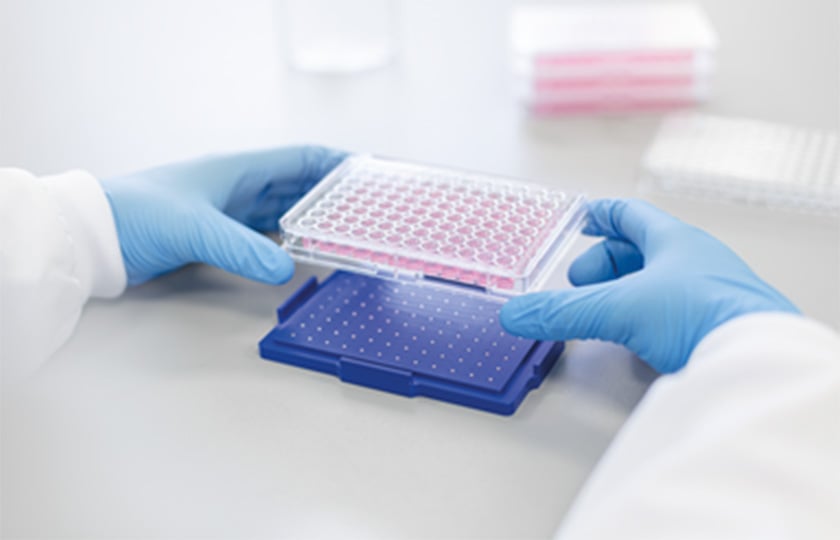
Reproducibility, standardization, miniaturization, scalability, and throughput will push cell culture technologies across the chasm. Magnetic 3D cell culture and bioprinting (M3D) are based on magnetizing cells using a biocompatible nanoparticle assembly NanoShuttle-PL consisting of poly-L-lysine, magnetic iron oxide, and gold nanoparticles that electrostatically adhere to cells [2]. When combined with flat-bottom cell-repellent microplates designed for HTS, magnetized cells can be easily manipulated with magnetic fields [3] to precisely and reproducibly enable:
• Xenogenic-free culture
• HTS with standard laboratory automation
• Scalability - 6 to 1536 well
• Miniaturization of culture
• Co-culture different cell types
• Positioning of cells for faster imaging
• Use of flat-bottom plates for high-resolution imaging
• Delineation of small imaging area
• Holding cells in place for media exchange or other downstream manipulation
• Use with automated plate washers
• Z' > 0.5 with chemiluminescence assays with 384 and 1,536 well formats
M3D is an innovative tool that can replicate the in vivo phenotypes in vitro, ideal for HTS compound response profiling in a rapid, miniaturized, and cost-effective manner [4].
Virneliz et al. applied magnetic 3D bioprinting (M3D) in the first large-scale screening effort completing HTS on over 150K molecules against primary pancreatic cancer cells. It is the first demonstration that a screening campaign of this magnitude using clinically relevant, ex vivo 3D pancreatic tumor models obtained directly from biopsy can be performed in the same manner as a conventional drug screening using 2D cell models [5].
Hou et al. also describe the utility of 3D magnetic cell culture for large-scale drug screening [6]. The researchers developed an HTS-compatible method to consistently produce organoids in standard 384 and 1536 well flat-bottomed plates by combining the use of a cell-repellent surface with magnetic force bioprinting technology. They validated this method by studying the effect of well-characterized anticancer agents against four patient-derived pancreatic cancer primary cells associated with KRAS mutations. The technology was tested for compatibility with HTS automation by performing a cytotoxicity pilot screening of ~3300 approved drugs [6].
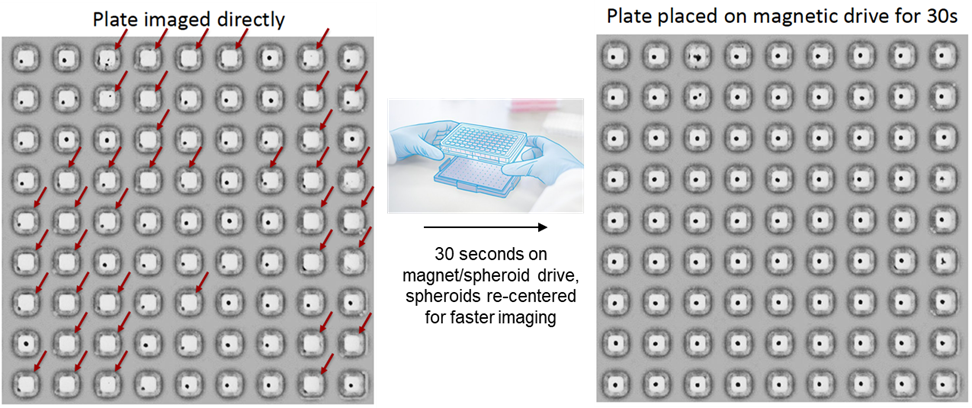
With M3D, magnetic fields can easily and precisely move and position spheroids to speed up imaging, which is ideal for a broad range of automation and HTS applications. Below, we show an example of how magnetic fields can precisely position spheroids at the center of the microwell for faster imaging.
More effective and faster imaging
Dots in the microwell are pictures of real spheroids in a flat bottom 384 well cell-repellent microplate. (Left) When using flat bottom plates, needed for high-resolution imaging, thermal motion within the well can move the spheroids off the center of the well (~ 25% well off center), which makes it challenging to acquire images. (Right)
Placing the flat bottom microwell plate on M3D magnetic drive for few seconds prior to imaging can center each spheroid back to the middle wells, allowing more effective and faster imaging.
Next, magnetic fields can also hold magnetized cultures in place, enabling easy media exchange with plate washers [7] and with more advanced tools like the BlueWasher from BlueCatBio [8].
What are the current 3D cell culture approaches for HTS?3D cell cultures can represent different organ models needed for HTS, including the specific phenotype, complex tissue signaling, and cellular interactions. There are several ways to produce 3D cell cultures, which can differ in the amount of manipulation, equipment, and reagents required. Bioprinting 3D culture systems Scaffold-free 3D culture systems Scaffold-based 3D culture systems |
A highly translational tool
M3D enabled HTS is a translational tools that can mimic the tumor microenvironment by recapitulating the complex genetic and molecular compositions of various tissue types. They can capture phenotypes that are predictive of in vivo biology needed to accelerate the identification of drug targets, streamline drug testing, and predict drug response in drug discovery and personalized medicine.
References
[1] Fang Y, Eglen RM. Three-Dimensional Cell Cultures in Drug Discovery and Development. SLAS Discov. 2017 Jun;22(5):456-472. doi: 10.1177/1087057117696795. Erratum in: SLAS Discov. 2021 Oct;26(9):NP1. PMID: 28520521; PMCID: PMC5448717.
[2] Tseng H, Souza GR. m3d cell culture: biocompatibility of Nanoshuttle-pl and the magnetic field. https://www.gbo.com/fileadmin/media/GBO-International/01_Downloads_BioScience/SALES_White_Papers/F075068_m3D_White_Paper_Biocompatibility_Nanoshuttle_EN.pdf
[3] Tseng H, Gage JA, Raphael RM, Moore RH, Killian TC, Grande-Allen KJ, Souza GR. Assembly of a three-dimensional multitype bronchiole coculture model using magnetic levitation. Tissue Eng Part C Methods. 2013 Sep;19(9):665-75. doi: 10.1089/ten.TEC.2012.0157. Epub 2013 Feb 25. PMID: 23301612.
[4] Rodboon T, Yodmuang S, Chaisuparat R, Ferreira JN. Development of high-throughput lacrimal gland organoid platforms for drug discovery in dry eye disease. SLAS Discov. 2022 Apr;27(3):151-158. doi: 10.1016/j.slasd.2021.11.002. Epub 2021 Dec 4. PMID: 35058190.
[5] Fernandez-Vega V, Hou S, Plenker D, Tiriac H, Baillargeon P, Shumate J, Scampavia L, Seldin J, Souza GR, Tuveson DA, Spicer TP. Lead identification using 3D models of pancreatic cancer. SLAS Discov. 2022 Apr;27(3):159-166. doi: 10.1016/j.slasd.2022.03.002. Epub 2022 Mar 17. PMID: 35306207.
[6] Hou S, Tiriac H, Sridharan BP, Scampavia L, Madoux F, Seldin J, Souza GR, Watson D, Tuveson D, Spicer TP. Advanced Development of Primary Pancreatic Organoid Tumor Models for High-Throughput Phenotypic Drug Screening. SLAS Discov. 2018 Jul;23(6):574-584. doi: 10.1177/2472555218766842. Epub 2018 Apr 19. PMID: 29673279; PMCID: PMC6013403.
[7] Larson B, Seldin J, Souza GR. Automated Walk-Away System to Perform Differentiation of 3D Mesenchymal Stem Cell. https://3dcellculture.gbo.com/wp-content/uploads/2019/03/Mesenchymal-Stem-Cells-Differentiation.pdf
[8] Hou S, Baillargeon P, Souza GR, Seldin J, Feist F, Scampavia L, Spicer TP. Validation of Magnetic 3D Spheroid Bioprinting in Combination with a BlueWasher. SLAS International Conference, 2018.
https://3dcellculture.gbo.com/wp-content/uploads/2019/03/2018-SLAS.pdf