Good imaging properties in microtiter plates are crucial for imaging and high-content screening. How well imaging ultimately works in this context depends on numerous factors that both interact and need to be optimized for individual research tasks. In this article, we briefly review imaging and high-content screening (HCS) applications and how imaging requirements determine microtiter plate characteristics for appropriate experimental results.
High-content screening – an emerging technology
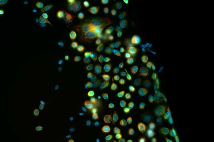
High-content screening (HCS) is one of the most important tools in advanced drug discovery. While classical high-throughput screening is occasionally based on rather simplistic readouts, HCS is a multiparametric version of cell based phenotypic screening and combines the technology of HTS with the advantages of automated imaging and image analysis. The high-resolution microscope-based techniques allow vast amounts of data to be obtained and analyzed using state-of-the-art (and sometimes AI-powered) image analysis software. The multitude of molecular features of individual cells that can be examined simultaneously are also the reason for the term "high content". The wealth of data generated by the HCS is both its strength and its weakness. The correct interpretation of data can still pose challenges to the scientific community. However, with the rapid progress in the field of artificial intelligence, the potential of this technology for drug development is expected to be greater than ever.
Simply put, cells are incubated with the substance to be screened, then the structures and molecular components of the cells are analyzed. Often proteins of interest are labeled with fluorescent dyes or chemiluminescence. Changes in cell phenotype or the quantitative presence or absence of the target proteins can then be measured with automated image analysis. Commercially available reporter assays are available and regularly used in HCS.
Imaging with fluorescence and bioluminescence
While the majority of HCS is fluorescence-based, bio- and chemiluminescence are popular for imaging because luminescence assays combine the advantages of high sensitivity, broad linearity, and robustness to library compounds and complex biological samples in a broader range of applications. The development of recombinant luciferase genes, enzymes, and substrates is providing further new detection opportunities. [1]
Multiplexing in high content screening
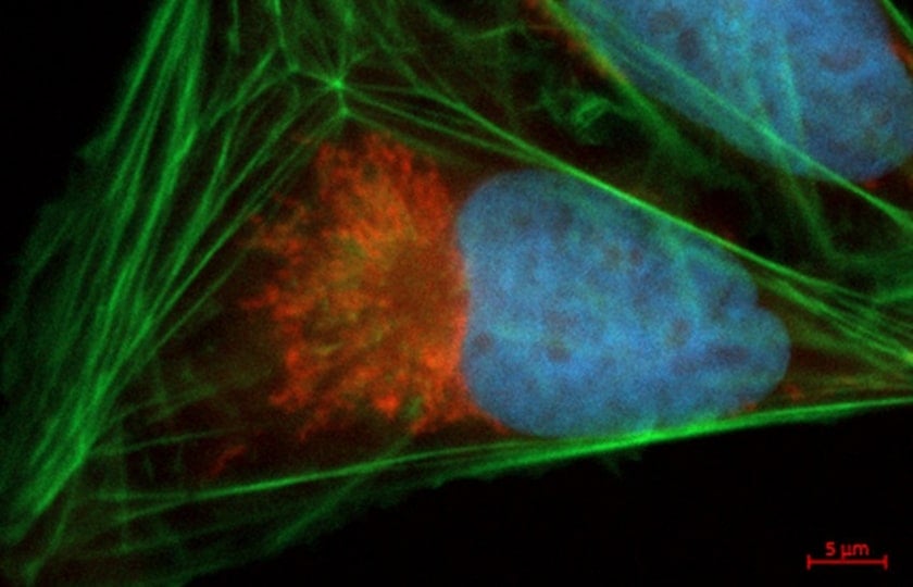
Multiple fluorescent dyes can be used simultaneously in cells and distinguished using optimal filter sets to generate multiparameter data sets or multiplex measurements. The fluorescent dyes are assembled to minimize spectral overlap so that the filter sets can separate the individual dyes. In addition, spectral deconvolution systems can also extract the fluorescence spectrum of each dye instead of separating them with filters.
Bioluminescence assays, multicolor HCS, and other applications all require specialized microtiter plates that meet the unique requirements of these techniques [2–4]. In the next blog post in this series, we will specifically address the details you need to consider when selecting microtiter plates for high content screening.
Would you like to learn more about the selection of optimal plastics labware for high-throughput screening with a special focus on imaging, and receive useful tips and tricks along the way? Don't miss any important updates and subscribe to our blog series now!
References
[1] Fan F, Wood KV. Bioluminescent assays for high-throughput screening. Assay Drug Dev Technol. 2007 Feb;5(1):127-36. doi: 10.1089/adt.2006.053. PMID: 17355205.
[2] Plastic Labware for Optimal Results in Modern Life Science Microscopy Using ZEISS Axio Observer and ZEISS Celldiscoverer 7. Technology Note. Philipp Wachter (Greiner Bio-One GmbH, Frickenhausen, Germany), Martin Gleisner, Volker Doering (Carl Zeiss Microscopy GmbH, Jena, Germany). February 2022 https://www.gbo.com/fileadmin/media/GBO-International/01_Downloads_BioScience/SALES_Application_Notes/Application_Note_GBO_labware_on_ZEISS_microscopes_EN.pdf
[3] SCREENSTAR: A new 1536 Well Microplate for High Content and High Throughput Screening. Greiner Bio-One forum – Technical Notes and Applications for Laboratory Work No. 15 https://www.gbo.com/fileadmin/media/GBO-International/01_Downloads_BioScience/SALES_Scientific_Publications/F073120_Forum_No._15_SCREENSTAR.pdf
[4] SCREENSTAR and CELLview: Microplates for Advanced Microscopy. Greiner Bio-One forum – Technical Notes and Applications for Laboratory Work No. 18 https://www.gbo.com/fileadmin/media/GBO-International/01_Downloads_BioScience/SALES_Scientific_Publications/F073787_Forum_No._18_SCREENSTAR_CELLview.pdf