Research to Viral Respiratory Infections Needs a Realistic Model
The need for greater insight into the mechanisms involved in respiratory diseases and their treatment and prevention has powered the development of increasingly realistic model cell-based systems. This includes a transition from simple single-layer cultures to differentiated, organotypic airway models with an air-liquid interface (ALI) and polarized epithelial cells.
The search for relevant cell models for respiratory infection
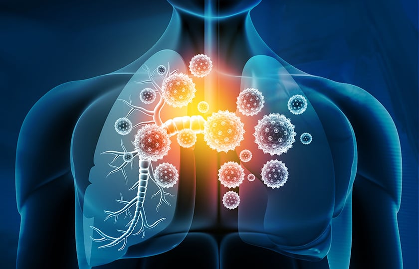
The respiratory system provides a sophisticated barrier that protects the body from invasion, but pathogens such as influenza, tuberculosis, rhinovirus, and coronaviruses have evolved and adapted to evade defense mechanisms. One important factor in the development of methods to prevent and treat respiratory infectious diseases is the availability of reliable and predictive model systems in the study of pathways and mechanisms involved in infection and replication.
Model systems range from animal models to in vitro lung cell models(1). Animal models have been routinely used since in vitro models lack one or another aspect of lung anatomy or physiology, but even animal models lack functional homology with the human respiratory system. There is also the issue of ethics, with major efforts being made to reduce the use of animals in research and align to the 3Rs initiative(2). This has led to intense efforts to develop and refine cell-based models.
Key factors to consider in a cell-based model system for the study of respiratory infection caused by pathogens such as coronavirus include:
1. Relevance to human respiratory cells:
The model system should use human respiratory cells or cell lines that closely mimic the anatomy, physiology, and immune responses of the human respiratory system. This is important for understanding how the virus interacts with the respiratory system and how the body responds to infection.
2. Viral strain and dose:
The model system should use a viral strain and dose that is relevant to the human infection. This is important for ensuring that the results of the study are applicable to human disease.
3. Reproducibility:
The model system should be reproducible and yield consistent results across different experiments and researchers. This is important for ensuring the reliability of the findings.
4. Availability and cost:
The model system should be easily accessible and cost-effective to use. This is important for making the model system widely available to researchers and reducing barriers to research.
Model refinement drives a transition from 2D to 3D and air-liquid interface culture
Submerged monolayer cultures derived from human respiratory tissues, such as normal human bronchial epithelial cells have been used widely for respiratory research and toxicology studies. The mammalian respiratory system consists of numerous specialized cell types, however, and such cultures cannot reproduce the differentiated phenotype of in vivo airway epithelial tissues. This has prompted the development of culture methods that generate well-differentiated 3D human airway organoids from, for example, human cell induced pluripotent stem cells. But while these organoids can be maintained under submerged conditions in long-term culture their spherical form and lack of an air interface limits their value when it comes to studying respiratory infections.
An alternative is to culture primary airway cells on microporous membrane scaffolds at the air-liquid interface (ALI) (1, 3–7). These differentiated, organotypic airway models based on air-liquid interface culture have several advantages compared to simpler models such as submerged or organoid cultures, including:
- They mimic in vivo structures, such as polarized epithelial cells, and metabolic functions more closely.
- They can be dosed in a more relevant manner.
- Primary cells that undergo cellular differentiation can reproduce an in vivo–like transcriptional profile similar to that of the human airway epithelium. This is important since infection by human respiratory pathogens, such as Bordetella pertussis, influenzae, and coronavirus, is highly dependent on the differentiation state of airway epithelium.
One important factor in the development of methods to prevent and treat respiratory infectious diseases is the availability of reliable and predictive model systems.
Organotypic ALI airway culture models for a range of applications
Cell models including ALI have been prepared from a wide range of primary cells and cell lines from nasal, bronchial, and alveolar epithelium(5, 6). There has been a significant increase in the use of polarized epithelial cells grown on permeable membranes(5) and the physiological relevance of the in vitro models can also be increased by co-culturing(7). Modelling can achieve a high level of mimicry. For example, human respiratory cells can be grown in air-liquid interface as organotypic airway cultures with polarized epithelial cells to resemble the human airway both morphologically (basal, ciliated, and secretory cells) and functionally (beating cilia and mucus secretion)(8).
Applications for cell models with ALI range from the study of respiratory pathogens to diseases such as asthma, cystic fibrosis, lung cancer, and chronic obstructive pulmonary disease (COPD)(7).
ALI models can also be used to screen for therapeutically relevant agents and their effectiveness on virus inactivation(7). For example, SARS-CoV-2 could be isolated from bronchial lavage fluid of patients suffering from COVID-19 disease and studied using ALI models of primary human bronchial epithelial cells. As a result, polarized ALI cultures proved their value as a high–throughput tool for rapidly gaining information about infection, replication, and pathogenesis of the virus.
In the next article in this series, we will look at a specific example of how introducing an air-liquid interface can transform the value of cell cultures to generate relevant data for respiratory infections.
Ready to enter the next level?
Please fill out this form and contact our experts today to find the perfect solution for you!
Don't miss our regular updates on scientific topics around 3D Cell Culture
References
[1] Cao X, Coyle JP, Xiong R, Wang Y, Heflich RH, Ren B, Gwinn WM, Hayden P, Rojanasakul L. Invited review: human air-liquid-interface organotypic airway tissue models derived from primary tracheobronchial epithelial cells-overview and perspectives. In Vitro Cell Dev Biol Anim. 2021 Feb;57(2):104-132. doi: 10.1007/s11626-020-00517-7. Epub 2020 Nov 11. PMID: 33175307; PMCID: PMC7657088.
[2]Russell WMS, Burch RL. 1959. (as reprinted 1992). The principles of humane experimental technique. Wheathampstead (UK): Universities Federation for Animal Welfare.
[3]Adler KB, Schwarz JE, Whitcutt MJ, Wu R. A new chamber system for maintaining differentiated Guinea pig respiratory epithelial cells between air and liquid phases. Biotechniques 1987; 5:462–465
[4]Whitcutt MJ, Adlet KB, Wu R. A biphasic chamber system for maintaining polarity of differentiation of cultured respiratory tract epithelial cells. In Vitro Cell Dev Biol. 1988; 24:420–428
[5]Hasan S, Sebo P, Osicka R. A guide to polarized airway epithelial models for studies of host-pathogen interactions. FEBS J. 2018 Dec;285(23):4343-4358. doi: 10.1111/febs.14582. Epub 2018 Jul 2. PMID: 29896776.
[6]Silva S, Bicker J, Falcão A, Fortuna A. Air-liquid interface (ALI) impact on different respiratory cell cultures. Eur J Pharm Biopharm. 2023 Mar;184:62-82. doi: 10.1016/j.ejpb.2023.01.013. Epub 2023 Jan 22. PMID: 36696943.
[7] Baldassi D, Gabold B, Merkel O. Air-liquid interface cultures of the healthy and diseased human respiratory tract: promises, challenges and future directions. Adv Nanobiomed Res. 2021 May 6;1(6):2000111. doi: 10.1002/anbr.202000111. PMID: 34345878; PMCID: PMC7611446.
[8] Levardon H, Yonker LM, Hurley BP, Mou H. Expansion of Airway Basal Cells and Generation of Polarized Epithelium. Bio Protoc. 2018 Jun 5;8(11):e2877. doi: 10.21769/BioProtoc.2877. PMID: 30009215; PMCID: PMC6040659.